
Dental Implant CBCT Planning: What Patients Should Notice
Learn what implant clinicians evaluate on CBCT, including bone height, ridge width, sinus floor, mandibular canal, mental foramen, and surgical guides.
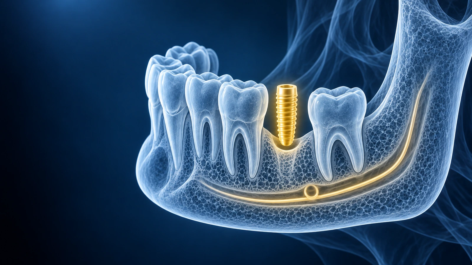
Implant CBCT planning is about fitting a restoration into real anatomy. The scan helps clinicians evaluate whether there is enough bone, whether a graft may be needed, and how close important structures are to the planned implant path.
Patients should focus on understanding the plan, not picking an implant size. The important questions are why the proposed position works, what risks exist, and whether alternatives have been considered.
What Implant Clinicians Measure
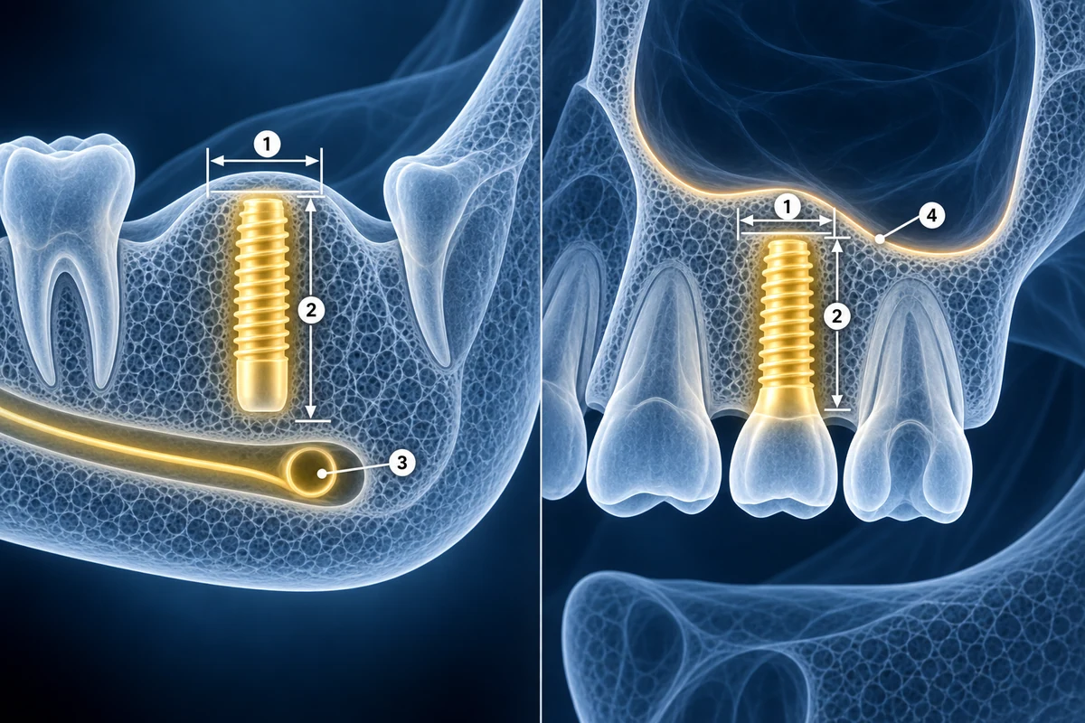
- Ridge width and available bone height at the proposed site
- Distance to mandibular canal, mental foramen, sinus floor, or nasal floor
- Socket healing, graft volume, bone contour, and cortical boundaries
- Prosthetic direction, neighboring roots, and surgical guide planning
Understand the Scan Before Consent
Open your CBCT privately, then ask your implant clinician how the scan changed the placement, grafting, or guide plan.
Read Implant Planning GuideKey Takeaways
- CBCT supports implant planning by showing bone in 3D
- Bone dimensions are only part of the implant decision
- Sinus and nerve distances are core safety questions
- Final planning must be done by a licensed dental clinician
Frequently Asked Questions
Does every implant need CBCT?
Many implant workflows use CBCT because it shows 3D bone and anatomy, but the need depends on the case, site, local standards, and clinician judgment.
Can CBCT show if bone grafting will work?
CBCT can show current bone dimensions and defects. It cannot guarantee graft success, which depends on biology, technique, healing, smoking status, systemic health, and follow-up care.
Related Articles
Learn how to understand dental CBCT views, slices, MPR planes, tooth roots, jawbone, sinuses, mandibular canal, and common report terms.
Compare dental CBCT and panoramic X-ray for wisdom teeth, implants, jaw cysts, root problems, orthodontics, radiation tradeoffs, and second opinions.
Related Conditions
Ready to analyze your imaging? Upload your MRI or X-ray for AI-powered analysis
Upload your MRI or X-ray DICOM files for private, AI-powered analysis. 4 models analyze independently — raw scan files stay in your browser.
Start AnalysisMedical Disclaimer: This page is for informational and educational purposes only. It does not constitute medical advice, diagnosis, or treatment. AI-generated analysis may contain errors. Always consult a qualified healthcare professional for medical decisions. Full Disclaimer