
Common Symptoms
- Jaw joint pain, clicking, locking, or limited opening
- Question about condyle flattening, erosion, remodeling, or asymmetry
- History of arthritis, trauma, bite change, or facial asymmetry
- Need to distinguish bony CBCT findings from soft-tissue MRI questions
TMJ CBCT Jaw Joint Findings Explained
View jaw and TMJ CBCT scans online and understand condyle shape, joint space, erosions, flattening, remodeling, and when MRI is needed for disc or soft tissue questions.
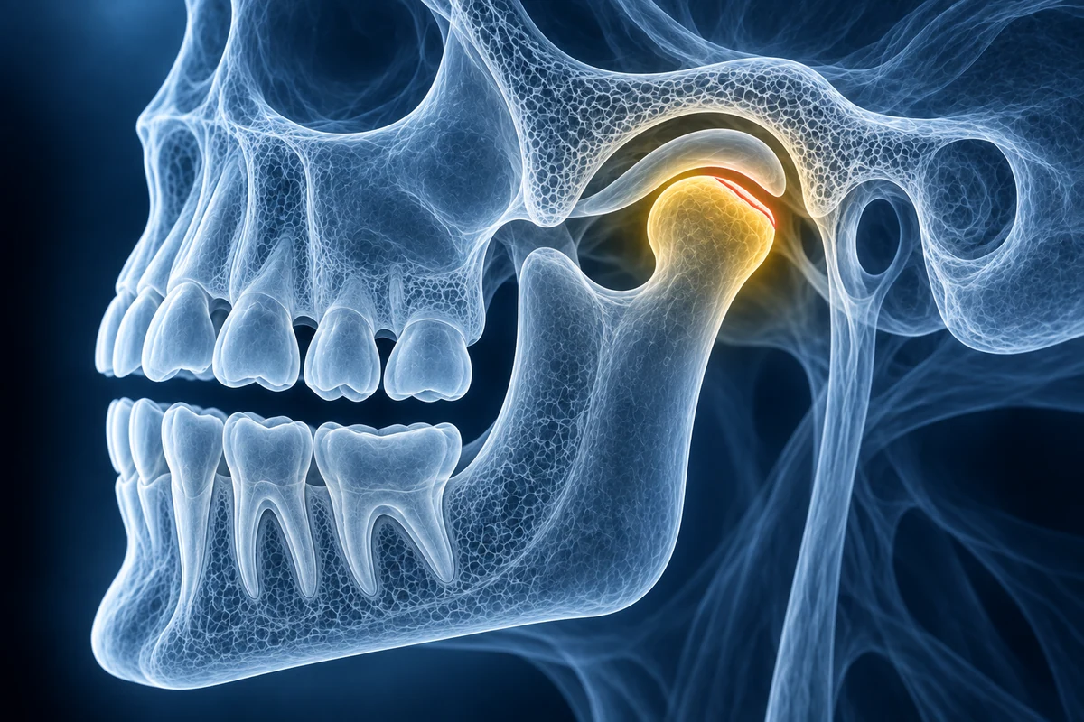
TMJ CBCT shows the bony parts of the jaw joint, especially the mandibular condyle and temporal bone. It can help visualize flattening, erosion, osteophytes, remodeling, and joint space asymmetry.
CBCT does not show the articular disc as well as MRI. If the main question is disc displacement, inflammation, or soft tissue, your clinician may recommend MRI instead of, or in addition to, CBCT.
For jaw and TMJ CBCT, keep the question dental and bony: condyle shape, joint space, erosion, remodeling, and asymmetry. The dental CBCT reading guide explains slice review, while CBCT vs panoramic X-ray covers when 3D depth is useful for jaw findings.
TMJ CBCT Findings to Understand
- Condylar flattening, erosion, sclerosis, osteophytes, or bony remodeling
- Joint space asymmetry and condyle position in the fossa
- Degenerative changes compared with the opposite jaw joint
- Limits of CBCT for disc position and inflammatory soft tissue findings
Review TMJ Bone Anatomy in 3D
Open the CBCT scan in your browser, compare both joints, and bring clearer questions to your dentist, oral surgeon, or TMJ specialist.
Open TMJ CBCTFrequently Asked Questions
Is CBCT or MRI better for TMJ pain?
CBCT is better for bony anatomy. MRI is better for the disc, inflammation, and soft tissues. The best test depends on whether your clinician is asking a bone question or a disc and soft-tissue question.
Can CBCT show a displaced TMJ disc?
Usually not reliably. Disc position is a soft-tissue question and is better evaluated with MRI. CBCT can show secondary bony changes that may accompany chronic joint problems.
Do bony TMJ changes always explain pain?
No. Imaging findings and symptoms do not always match. TMJ pain can involve muscle, disc, bite, inflammation, stress, and parafunction factors, so clinical evaluation remains essential.
Related Articles
📖
Do You Need a 3T MRI for Dental or Jaw Imaging?
📖Learn when dental and jaw problems usually need CBCT, panoramic X-ray, or intraoral imaging instead of 3T MRI, and when TMJ or soft-tissue MRI may make sense.
CBCT vs Panoramic X-ray: When Dental 3D Imaging Helps
Compare dental CBCT and panoramic X-ray for wisdom teeth, implants, jaw cysts, root problems, orthodontics, radiation tradeoffs, and second opinions.
Upload your imaging to check for TMJ CBCT
Upload your MRI or X-ray DICOM files for private, AI-powered analysis. 4 models analyze independently — raw scan files stay in your browser.
Upload & AnalyzeMedical Disclaimer: This page is for informational and educational purposes only. It does not constitute medical advice, diagnosis, or treatment. AI-generated analysis may contain errors. Always consult a qualified healthcare professional for medical decisions. Full Disclaimer