
Wisdom Tooth CBCT Guide: Roots, Nerves, and Sinus Anatomy
Understand how CBCT helps evaluate impacted wisdom teeth, root shape, mandibular canal proximity, maxillary sinus relationship, and extraction planning questions.
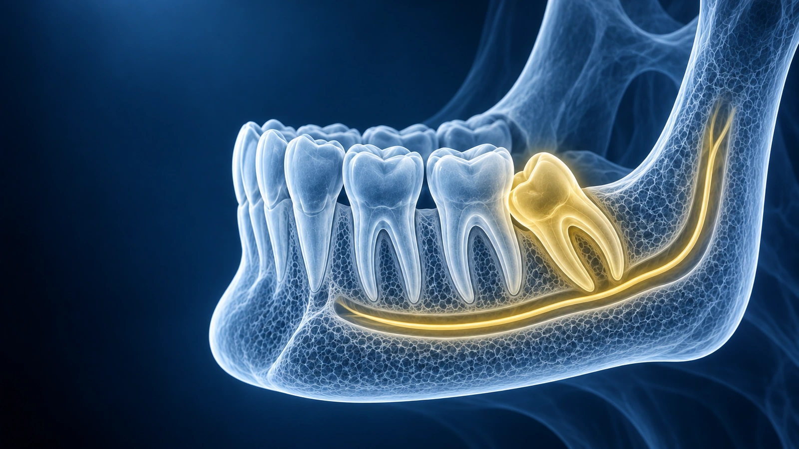
Wisdom tooth CBCT helps show the 3D relationship between a third molar, its roots, jawbone, mandibular canal, and maxillary sinus. This is especially useful when a panoramic X-ray suggests close nerve or sinus proximity.
The scan does not decide whether removal is required. It helps your oral surgeon understand anatomy and explain treatment options, risks, and alternatives.
Third molars are the last teeth to form, and they often finish developing after the jaw has run out of room for them. When that happens the tooth stops part-way through its eruption and settles at whatever angle the surrounding bone and neighbouring roots allow. The result is an impaction, and its angle is not a cosmetic detail: it decides which direction the tooth would have to travel to come out, how much bone sits in the way, and whether the crown is pressing against the second molar behind it.
Everything a cone beam scan adds comes back to one limitation of a flat film. A panoramic image compresses a curved jaw onto a single plane, so two structures that are centimetres apart in the cheek-to-tongue direction land on top of one another in the picture. Cone beam imaging restores that missing third dimension. This page explains what that extra dimension is used for and how the findings are usually described; it is general education and not a diagnosis or a treatment recommendation.
How Impaction Is Classified
The first descriptor is angulation, measured against the long axis of the second molar. A mesioangular tooth leans forward toward the front of the mouth, a distoangular tooth tips backward, a vertical tooth stands upright but has not fully erupted, and a horizontal tooth lies on its side with the crown facing the neighbouring root. Each angle presents a different face of the tooth to the surgeon and a different amount of overlying bone.
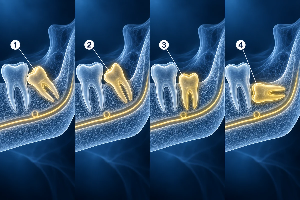
A second descriptor records depth and available space. Depth is expressed by comparing the height of the impacted crown with the crown of the second molar — level with it, partly below it, or completely below it. Available space compares the width of the crown with the room between the second molar and the front edge of the ascending part of the jaw. Together the two descriptors tell you whether the obstacle is bone above the tooth, a lack of room behind it, or both.
Root form is the third element and the one flat images describe least reliably. Reports note how many roots there are, whether they are fused into a single cone or splayed apart, whether any root is hooked or curved, and whether a root wraps around the nerve canal rather than merely touching it. Because a cone beam volume can be rotated, these shapes are read directly rather than inferred.
What Each Scan Shows and What It Misses
A panoramic film remains the sensible first study. It surveys both jaws, all four third molars, and the surrounding bone in a single low-dose exposure, and for a great many wisdom teeth it answers every question that needs answering. It also carries a well-established set of warning signs that flag a tooth worth looking at more closely: darkening of the root where it crosses the canal, loss of one or both of the white lines that outline the canal, a bend in the canal as it passes the tooth, and narrowing of either the root or the canal at the crossing point.
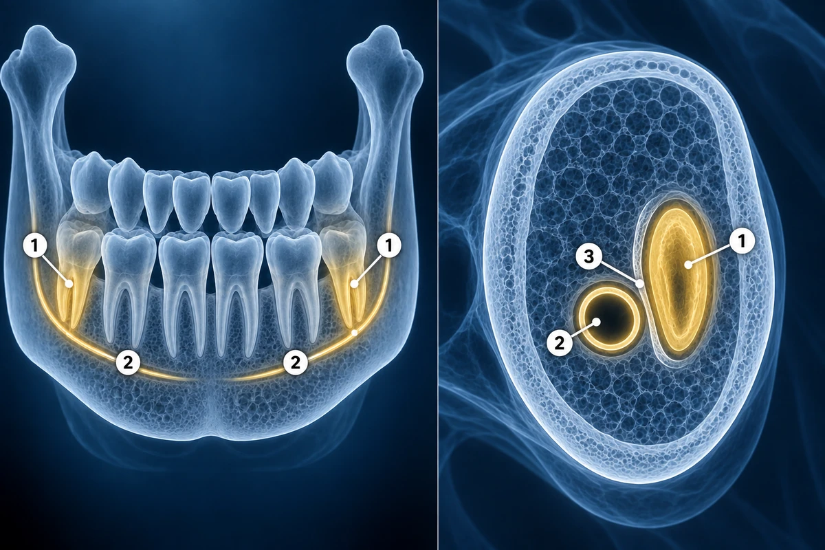
What the panoramic film cannot do is tell you where the canal sits in the cheek-to-tongue direction. A canal that appears to cross a root may in fact pass well to one side of it, and one that appears clear may be pressed against the root surface. The flat image also distorts vertical and horizontal proportions unevenly depending on how the patient was positioned, so measurements taken from it are approximations.
Cone beam imaging resolves exactly that ambiguity. Cross-sectional slices show whether the canal runs to the cheek side, the tongue side, directly below, or between splayed roots, and whether a thin shell of cortical bone still separates the two. It also shows the shape of the sinus floor above an upper wisdom tooth and any bone loss or cyst formation around the crown. Its limits are real as well: the radiation dose is higher than a panoramic film, metal restorations nearby can streak the images, and it shows only hard tissue, so the nerve itself is never visualised — only the tunnel it travels in.
Report Phrases and What They Mean
- "Loss of the cortical boundary of the canal" — the thin white shell between root and canal is no longer visible at the point they meet
- "Canal displaced lingually by the root apex" — the tunnel has been pushed toward the tongue side rather than simply crossing the root
- "Grooving of the root by the canal" — the canal has indented the root surface, describing intimate contact without penetration
- "Roots project into the sinus with a dehiscent floor" — an upper molar root reaches the sinus and the bony floor above it is incomplete
- "Pericoronal radiolucency exceeding normal follicular width" — the dark zone around the crown is wider than an expected developmental space
- "Distal bone loss at the adjacent second molar" — bone has been lost on the side of the neighbouring tooth that faces the impacted one
Key Questions to Ask
- Are the roots touching or surrounding the mandibular canal?
- Is there a visible cortical boundary between the tooth and canal?
- For upper wisdom teeth, how close are the roots to the sinus floor?
- Does the scan change the extraction plan, timing, or alternatives?
What Usually Happens Next
A scan that shows a clear separation between root and canal generally does not change anything — the case proceeds as the panoramic film already suggested. The scan earns its place when it reveals a relationship the flat image could not resolve, because that is the situation where the surgical approach, the sectioning of the tooth, or the choice to leave the roots in place may be planned differently. Which of those applies to you is a conversation for your oral surgeon.
Not every impacted wisdom tooth is destined for removal. A tooth that is fully covered by bone, causing no symptoms, showing no bone loss at the neighbouring molar, and not interfering with anything else may reasonably be watched. Monitoring means periodic clinical review rather than repeated scanning, and the reasons to revisit the decision are usually symptoms, recurrent inflammation around a partly erupted crown, or changes seen on a routine film.
Repeat cone beam imaging is uncommon and is not part of routine follow-up. It is considered when something has changed that the earlier study cannot answer — a pericoronal area that has enlarged on a later panoramic film, a retained root fragment whose position needs defining, or a planned second procedure where the anatomy has altered since the first scan. Because the dose is higher than a flat film, each request is expected to be justified by a question the existing images cannot settle.
Open the Wisdom Tooth CBCT
Use the viewer first, then read the condition guide for impacted wisdom teeth.
Read Impacted Wisdom Tooth GuideKey Takeaways
- CBCT clarifies depth relationships that panoramic X-rays flatten
- The mandibular canal is the key structure for lower wisdom teeth
- Sinus proximity matters for upper wisdom teeth
- Surgical risk discussion belongs with an oral surgeon
- Impaction is described by angulation, by depth relative to the second molar, and by the space available behind it
- Panoramic warning signs — root darkening, interrupted canal outlines, canal diversion, narrowing at the crossing — are what usually prompt a cone beam request
- Cone beam imaging shows the canal on the cheek or tongue side of the root, which a flat film cannot resolve
- The scan shows the bony canal, never the nerve itself, and carries a higher dose than a panoramic film
Frequently Asked Questions
Does close nerve proximity mean I will have numbness?
No. Close proximity is a risk factor, not a prediction. Your surgeon evaluates the exact anatomy, symptoms, and treatment options before counseling you.
Can CBCT show whether coronectomy is an option?
CBCT can inform that discussion by showing root and canal relationships, but coronectomy suitability depends on clinical judgment and case-specific factors.
Why can a panoramic X-ray not answer the nerve question by itself?
A panoramic image flattens a curved jaw into one plane, so everything along the cheek-to-tongue direction is stacked into a single shadow. A root and a canal that appear to overlap on that image may be separated by several millimetres of bone, and two teeth with identical-looking films can have entirely different anatomy. The flat film is very good at telling you which teeth deserve a closer look, and the recognised warning signs exist for exactly that purpose, but it cannot state which side of the root the canal passes. Whether that distinction matters in your case is for your clinician to judge.
What does the angle of the tooth change?
The angle determines which direction the tooth would need to move to leave its socket and what is standing in the way. A tooth leaning forward has its crown pressed against the neighbouring molar, so that contact point becomes the focus. A tooth lying on its side presents its crown sideways and sits under a different thickness of bone. A tooth tipped backward points toward the ascending part of the jaw, where access is more restricted. Reports describe the angle because it shapes the plan, not because one angle is inherently worse than another.
Should every wisdom tooth be scanned before removal?
No, and the guiding principle is that imaging should be justified by a question that will change what is done. Many wisdom teeth are well away from the canal and the sinus, and a panoramic film supplies everything needed. Cone beam imaging involves a higher radiation dose than a flat film, so it is generally reserved for cases where the relationship to the canal or sinus is genuinely uncertain, where earlier images show warning signs, or where a specific surgical decision depends on the answer. Your dentist or oral surgeon decides whether your case meets that bar.
Can the scan show the nerve itself?
Not directly. Cone beam imaging renders hard tissue, so what you see is the bony tunnel through the lower jaw, not the nerve bundle travelling inside it. The assumption that the nerve follows the tunnel is reasonable and is what planning is based on, but the canal outline can be faint or incomplete in some people, and small accessory branches are not always traceable. This is one of several reasons the scan informs a surgical plan rather than replacing the surgeon's judgement during the procedure.
Related Articles
Learn how to understand dental CBCT views, slices, MPR planes, tooth roots, jawbone, sinuses, mandibular canal, and common report terms.
Compare dental CBCT and panoramic X-ray for wisdom teeth, implants, jaw cysts, root problems, orthodontics, radiation tradeoffs, and second opinions.
Related Conditions
Ready to analyze your imaging? Upload your MRI or X-ray for AI-powered analysis
Upload your MRI or X-ray DICOM files for private, AI-powered analysis. 4 models analyze independently — raw scan files stay in your browser.
Start AnalysisMedical Disclaimer: This page is for informational and educational purposes only. It does not constitute medical advice, diagnosis, or treatment. AI-generated analysis may contain errors. Always consult a qualified healthcare professional for medical decisions. Full Disclaimer