
Root Fracture CBCT Signs: What Dental CT Can and Cannot Show
Understand possible root fracture signs on CBCT, metal artifact limits, vertical bone defects, fracture lines, and why clinical testing still matters.
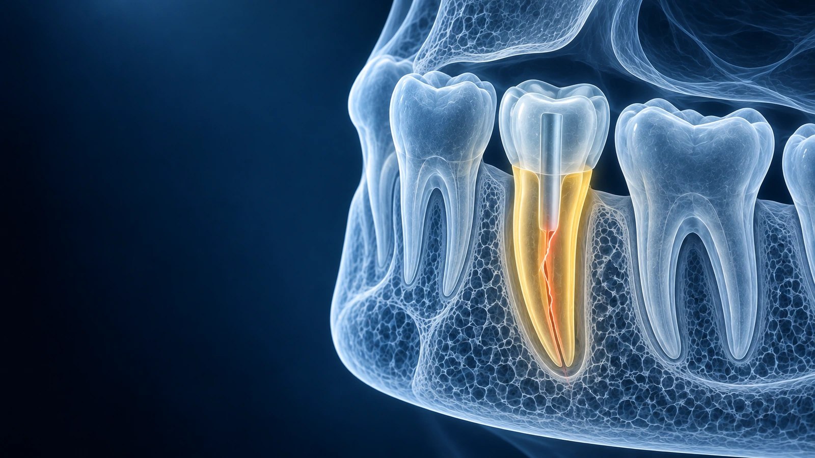
Root fractures are challenging because the line may be tiny, hidden by metal artifact, or visible only in one plane. CBCT can help by separating overlapping anatomy, but it cannot replace clinical dental testing.
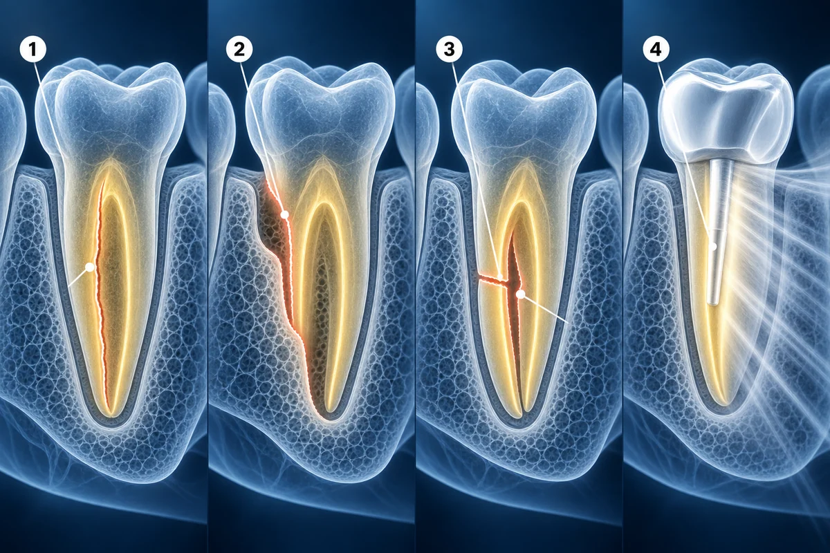
A clinician usually looks for the fracture line, an isolated vertical bone defect, root separation, symptoms when biting, periodontal probing changes, and the history of root canal treatment, post placement, crown placement, or trauma.
CBCT Signs That May Be Discussed
- Thin radiolucent line crossing or tracking along a root
- Localized vertical bone loss next to one root surface
- Root segment separation, unusual root shape, or adjacent lesion
- Metal artifact that limits confidence around posts or crowns
Review the Suspected Root from Several Angles
Open the CBCT and then use the root fracture guide to prepare the right clinical questions.
Read Root Fracture GuideKey Takeaways
- CBCT can help but does not prove every root fracture
- Metal artifact is a major limitation
- Clinical symptoms and probing findings matter
- Restorability determines many treatment decisions
Frequently Asked Questions
Can a cracked root be saved?
Some cracks are manageable, while many vertical root fractures have poor prognosis. The answer depends on fracture location, symptoms, periodontal support, and whether the tooth is restorable.
Why might my CBCT report say suspected fracture?
Imaging may show indirect signs without a definite fracture line. Dentists often use the word suspected when scan quality, artifact, or clinical uncertainty remains.
Related Articles
Learn how to understand dental CBCT views, slices, MPR planes, tooth roots, jawbone, sinuses, mandibular canal, and common report terms.
Plain-language guide to periapical lesions on CBCT, including root tip radiolucency, sinus relationship, cortical bone change, and endodontic follow-up.
Related Conditions
Ready to analyze your imaging? Upload your MRI or X-ray for AI-powered analysis
Upload your MRI or X-ray DICOM files for private, AI-powered analysis. 4 models analyze independently — raw scan files stay in your browser.
Start AnalysisMedical Disclaimer: This page is for informational and educational purposes only. It does not constitute medical advice, diagnosis, or treatment. AI-generated analysis may contain errors. Always consult a qualified healthcare professional for medical decisions. Full Disclaimer