
Periapical Lesion on CBCT: What a Root Tip Finding Means
Plain-language guide to periapical lesions on CBCT, including root tip radiolucency, sinus relationship, cortical bone change, and endodontic follow-up.
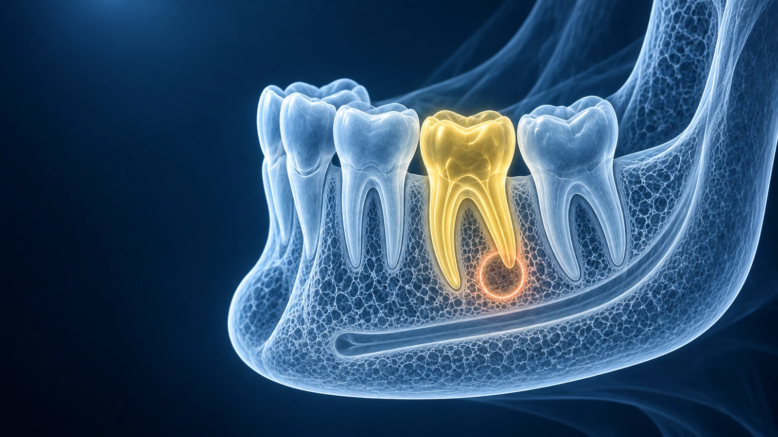
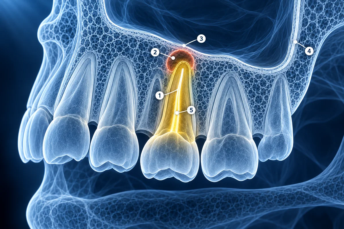
A periapical lesion is a finding around a tooth root tip. It may be connected to pulp infection, inflammation, previous treatment, trauma, or another dental condition. CBCT helps show its location and extent, but clinical testing explains what it means.
Useful details include the involved tooth, the root or canal involved, size, sinus relationship, cortical bone changes, and whether there is existing root canal filling or restoration.
Questions for an Endodontic Visit
- Which tooth and root is the lesion centered on?
- Does the tooth test vital, previously treated, or symptomatic?
- Is the sinus floor, nasal floor, or cortical bone affected?
- Are root canal treatment, retreatment, surgery, extraction, or monitoring being considered?
Match the Scan to the Right Tooth
Use the periapical lesion condition guide to connect the CBCT finding with practical follow-up questions.
Read Periapical Lesion GuideKey Takeaways
- CBCT localizes root tip findings in 3D
- Imaging does not replace pulp testing and dental examination
- Sinus and cortical bone relationships can affect treatment planning
- Existing dental work can change interpretation
Frequently Asked Questions
Can a periapical lesion heal?
Some lesions can resolve after appropriate dental treatment, but healing depends on the cause, tooth restorability, infection control, and follow-up. Your dentist or endodontist should guide monitoring.
Why is CBCT used if I already had a small dental X-ray?
A small X-ray can flatten overlapping anatomy. CBCT can show the lesion in depth and clarify sinus, root, canal, or cortical bone relationships.
Related Articles
Learn how to understand dental CBCT views, slices, MPR planes, tooth roots, jawbone, sinuses, mandibular canal, and common report terms.
Understand possible root fracture signs on CBCT, metal artifact limits, vertical bone defects, fracture lines, and why clinical testing still matters.
Related Conditions
Ready to analyze your imaging? Upload your MRI or X-ray for AI-powered analysis
Upload your MRI or X-ray DICOM files for private, AI-powered analysis. 4 models analyze independently — raw scan files stay in your browser.
Start AnalysisMedical Disclaimer: This page is for informational and educational purposes only. It does not constitute medical advice, diagnosis, or treatment. AI-generated analysis may contain errors. Always consult a qualified healthcare professional for medical decisions. Full Disclaimer