
How to Read a Dental CBCT Scan: A Patient Guide
Learn how to understand dental CBCT views, slices, MPR planes, tooth roots, jawbone, sinuses, mandibular canal, and common report terms.
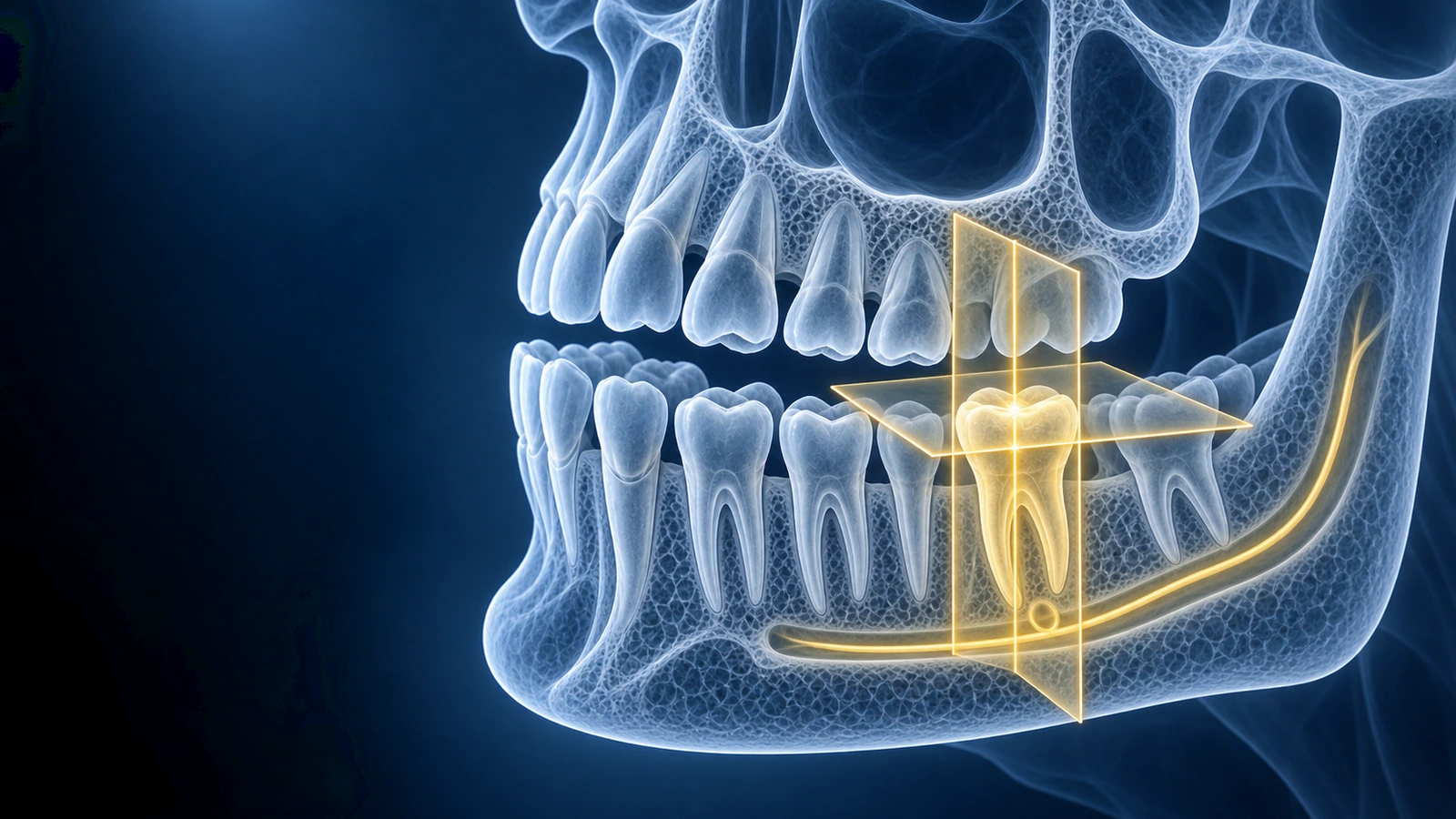
Dental CBCT is a three-dimensional scan. Instead of one flat image, it creates a volume that can be viewed as thin slices and reconstructed into axial, coronal, and sagittal planes. That depth is why CBCT is useful for wisdom teeth, implant planning, root findings, jaw lesions, and TMJ bone questions.
A patient does not need to interpret every detail to benefit from the scan. The goal is to understand the basic anatomy, identify the area your clinician is discussing, and prepare precise questions for a second opinion.
Core CBCT Views
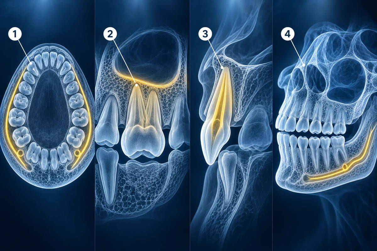
- Axial slices: useful for tooth roots, canals, cortical bone, and jaw width
- Coronal slices: useful for sinus floor, implant height, and vertical anatomy
- Sagittal slices: useful for tooth angulation, root length, and front-back depth
- 3D rendering: helpful for orientation, but treatment decisions rely on slices
What to Check Before Your Appointment
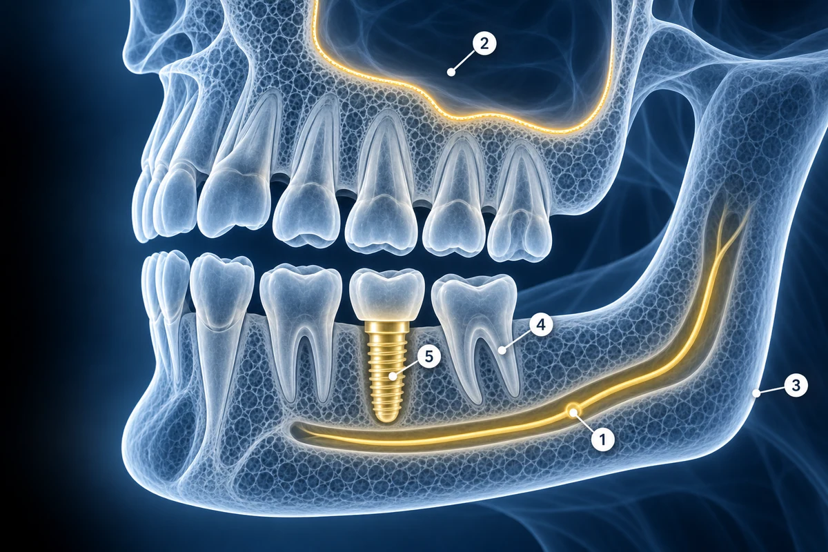
First, confirm which tooth or jaw region is being discussed. Then identify nearby structures: the mandibular canal in the lower jaw, the maxillary sinus in the upper jaw, cortical bone boundaries, neighboring roots, and any existing dental work such as implants, posts, crowns, or root canal fillings. Use the free CBCT viewer to inspect these relationships without uploading raw files.
Key Takeaways
- CBCT is 3D; panoramic X-ray is a 2D projection
- Slices matter more than 3D renderings for clinical decisions
- Mandibular canal, sinus floor, and cortical bone are key landmarks
- Use CBCT to ask better questions, not to self-diagnose
Frequently Asked Questions
Can I read a dental CBCT scan myself?
You can learn the orientation and recognize the area being discussed, but formal interpretation requires dental and radiology training. Use patient review as preparation for a professional discussion.
Why does my CBCT look different from the 3D model?
The 3D model is a rendering of voxel data. It is useful for orientation, but thin slices show fine details and are more important for diagnosis and planning.
Related Articles
Compare dental CBCT and panoramic X-ray for wisdom teeth, implants, jaw cysts, root problems, orthodontics, radiation tradeoffs, and second opinions.
Understand how CBCT helps evaluate impacted wisdom teeth, root shape, mandibular canal proximity, maxillary sinus relationship, and extraction planning questions.
Learn when dental and jaw problems usually need CBCT, panoramic X-ray, or intraoral imaging instead of 3T MRI, and when TMJ or soft-tissue MRI may make sense.
Related Conditions
Ready to analyze your imaging? Upload your MRI or X-ray for AI-powered analysis
Upload your MRI or X-ray DICOM files for private, AI-powered analysis. 4 models analyze independently — raw scan files stay in your browser.
Start AnalysisMedical Disclaimer: This page is for informational and educational purposes only. It does not constitute medical advice, diagnosis, or treatment. AI-generated analysis may contain errors. Always consult a qualified healthcare professional for medical decisions. Full Disclaimer